Collagen fibrils
Nanomechanical properties of the most abundant structural protein in vertebrates
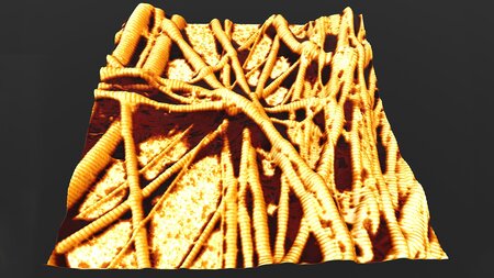
Atomic force microscopy image of collagen fibrils (orange, cross-striped) with adhering triacylglycerol molecules (dark areas). Image credit: Martin Dehnert and Robert Magerle.
Collagens are a major component of the connective tissue of vertebrates and provide mechanical strength to tissues. The most abundant type is type I collagen, which forms fibrils 30 to 300 nm in diameter and has a periodic structure with a lattice constant of 67 nm along the fibril axis. The amount and distribution of water, lipids (fat molecules), and molecular bonds between collagen molecules determine and control the mechanical properties of collagen fibrils. We discovered that collagen fibrils from chicken tendons contain an unexpectedly high amount of triacylglycerols, which a very common type of natural fat molecule. These fat molecules assemble between the collagen molecules and act as plasticizers, soften the collagen fibrils and reduce their water content. This finding is essential for understanding the biomechanics of connective tissue. Read more about our discovery in the university news and the original publication in Soft Matter [1].
We study the nanoscale mechanical properties of collagen fibrils using atomic force microscopy (AFM) and force spectroscopy. Through humidity control, we accurately control the water content of the fibrils. Using pointwise force–distance data, we reconstruct 3D depth profiles of the tip–sample interaction force, which allows us to distinguish the contributions of capillary force and adhesion from the viscoelastic properties of collagen fibrils [2,3]. This enables us to examine the nanomechanical properties of individual collagen fibrils in natural tendons with spatial resolution down to 10 nanometers [3,4].
The contact force and energy dissipation that occur when the tip interacts with the collagen fibril are independent of indentation velocity and can be more accurately described using a hysteresis model with return-point memory than using spring-dashpot models [4]. The EPICAL hysteresis model describes phenomenologically and in a unified form the sequences of mechanical phenomena that occur in AFM-based nanoindentation experiments: Elasto-plastic indentation, capillary adhesion and surface levelling (EPICAL). The model is based on force–distance data measured with the AFM during a large approach-retract cycle and predicts the force (output) and the dissipated energy for arbitrary indentation trajectories (input). An interactive input-output diagram of the EPICAL hysteresis model can be found here.
The swelling behavior of the fibrils provides direct information about the local content of free water molecules in the overlap and gap regions of the D band [5]. Using AFM-based nanotomography we imaged and reconstructed the spatial arrangement of individual collagen fibrils in bone [6].
References
[1] M. Dehnert, T. Klose, Y. Pan, D. R. T. Zahn, M. Voigtländer, J. F. Teichert, R. Magerle, Soft Matter (2025), DOI: https://doi.org/10.1039/D5SM00696A
[2] R. Magerle, M. Dehnert, D. Voigt, A. Bernstein, Anal. Chem. 92, 8741–8749 (2020).
[3] M. R. Uhlig, R. Magerle, Nanoscale 9, 1244–1256 (2017); accepted manuscript: arXiv:1910.00794.
[4] R. Magerle, P. Zech, M. Dehnert, A. Bendixen, A. Otto, Soft Matter 20, 2831–2839 (2024);
[5] E.-C. Spitzner, S. Röper, M. Zerson, A. Bernstein, R. Magerle, ACS Nano 9, 5683–5694 (2015).
[6] S. Röper, Dissertation, TU Chemnitz (2010). PDF