
optical density of states in photonic crystals
the group is using for the first time single CdSe/ZnS quantum dots to explore the local density of states in 3-dimensional photonic crystals. the above images show the defocusing patterns of the emission of single CdSe/ZnS quantum dots in a polystyrene opal. due to the anisotropic light propagation in the photonic crystal, the defocusing patterns are altered compared to a homogeneous material. this is the very first proof of a local measurement inside a 3-dimensional photonic crystal.


photonic crystals are new and unique materials which mimic the properties of semiconductors for photons. light is scattered by periodic dielectric structures causing an optical band structure, optical band gaps as well as optical defect states. therefore such photonic crystal structures are ideal materials for transporting and manipulating light as well as light emission by chromophores placed within a photonic crystal. the groups interest is this manipulation of light emission in photonic crystals which is studied with microscopic techniques including single molecule spectroscopy as well as theoretical aproaches.
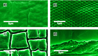
electron microscopy images of a colloidal photonic crystal made of polystyrene beads (260 nm)


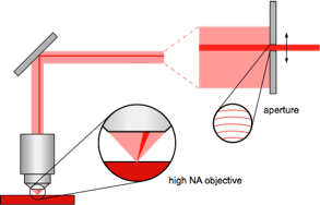
angle resolved microscopy scheme developed by the group to obtain angle resolved emission spectra
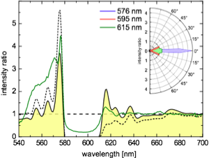
comparison of experimental and theoretical data concerning the fractional local density of states

the group has shown by theoretical calculations introducing a new quantity called fractional local density of states (FLDOS), that the local density of states is subject to an angular and spectral redistribution. the figure to the right shows the calculated ratio of rhodamine B emission spectra with and without a photonic crystal (solid line) together with the calculated FLDOS at various points at the unit cell. the strong emission enhancement at 575 nm is highly directive as diplayed in the polar plot.

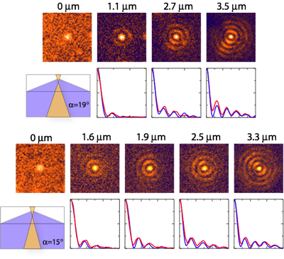

defocused emission patterns of single CdSe/ZnS quantum dots in a 3-dimensional photonic crystal. the graphs below the images show the a comparison of the experimental and simulated radial intensity distribution in the images. the simulation refer to a cone with no light propagation as depicted at the left side.
